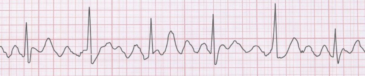
 Atrial Flutter ECG Example
Figure 1: Atrial Flutter - Characteristic ECG Pattern
Atrial Flutter ECG Example
Figure 1: Atrial Flutter - Characteristic ECG Pattern
🔑 Key Points at a Glance
- Heart Rate: Atrial 250-350 bpm, Ventricular variable
- Primary Significance: Organized atrial activity, stroke risk similar to AFib, highly ablatable
- Key Management: Rate control, consider cardioversion, catheter ablation (>90% success rate)
- Clinical Category: Clinical
Overview and Clinical Significance
Atrial Flutter represents an important cardiac rhythm pattern that clinicians must accurately identify. Organized atrial activity, stroke risk similar to AFib, highly ablatable
Understanding this rhythm is essential for emergency physicians, cardiologists, intensivists, and all healthcare providers involved in acute cardiac care. Early recognition and appropriate management can significantly impact patient outcomes.
ECG Characteristics and Recognition
📊 Diagnostic ECG Criteria
- Sawtooth flutter waves (F waves)
- Regular atrial rate ~300 bpm
- AV block (2:1, 3:1, 4:1)
- Negative flutter waves in II, III, aVF
Systematic ECG Analysis Approach
When analyzing any ECG, including suspected Atrial Flutter, follow this systematic approach:
- Rate: Calculate the ventricular rate using the 300-150-100-75-60-50 rule or count complexes in 6 seconds × 10
- Rhythm: Assess regularity by measuring R-R intervals across the strip
- P Waves: Identify presence, morphology, and relationship to QRS complexes
- PR Interval: Measure from start of P wave to start of QRS (normal: 0.12-0.20 seconds)
- QRS Complex: Assess duration (normal: 1mm is significant)
- T Waves: Check morphology, direction, and concordance with QRS
- QT Interval: Measure and correct for heart rate (QTc normal: Correlate ECG findings with clinical presentation - the patient, not the monitor, determines management urgency
Evidence-Based Management
Acute Management Strategy
Primary Treatment Approach: Rate control, consider cardioversion, catheter ablation (>90% success rate)
Pharmacologic Interventions
Rate control with beta-blockers or calcium channel blockers is typically first-line. Consider anticoagulation based on CHA₂DS₂-VASc score.
Procedural Considerations
Catheter ablation offers definitive cure with >90% success rate - consider referral for recurrent symptomatic episodes.
Differential Diagnosis
🔍 Consider These Mimics
- Atrial fibrillation - more irregular with no organized waves
- Atrial tachycardia - distinct P waves, not flutter waves
- AVNRT with 2:1 block - P waves buried in QRS
Complications and Risk Stratification
Potential complications associated with Atrial Flutter include:
- Thromboembolic events (stroke risk similar to AFib)
- Heart failure from rapid ventricular rates
- Tachycardia-induced cardiomyopathy
Long-Term Management and Follow-Up
Regular outpatient follow-up with cardiology or electrophysiology is recommended to monitor for progression and optimize therapy.
📅 Follow-Up Recommendations
- Cardiology follow-up within 2-4 weeks
- Consider Holter monitor or event recorder for recurrent symptoms
- Lifestyle modifications: exercise, stress reduction, avoid triggers
Common Pitfalls and How to Avoid Them
⚠️ Common Mistakes to Avoid
- Mistaking 2:1 flutter for sinus tachycardia - use carotid massage or adenosine to reveal flutter waves
- Cardioverting without anticoagulation - same stroke risk as AFib
- Missing opportunity for catheter ablation - highly effective cure
- Under-dosing rate control medications
Patient Education and Counseling
When counseling patients diagnosed with Atrial Flutter, address the following key points:
- Nature of the condition: Explain the rhythm abnormality in simple terms, avoiding medical jargon
- Prognosis: Provide realistic expectations about symptom control and quality of life
- Warning signs: Educate about symptoms requiring immediate medical attention (chest pain, syncope, severe dyspnea)
- Medication compliance: Importance of taking prescribed medications as directed
- Lifestyle modifications: Limit caffeine and alcohol, maintain healthy weight, exercise regularly (as tolerated), stress reduction
- Activity restrictions: Generally no restrictions once symptoms controlled
Evidence-Based Guidelines and References
Current management of Atrial Flutter is based on evidence from major clinical trials and consensus guidelines from professional societies including:
- American Heart Association (AHA) / American College of Cardiology (ACC) Guidelines
- European Society of Cardiology (ESC) Guidelines
- Advanced Cardiac Life Support (ACLS) Protocols
- Heart Rhythm Society (HRS) Expert Consensus Statements
📚 Level of Evidence
Most recommendations for acute management of Atrial Flutter are supported by Level B (limited randomized trials or observational studies) evidence.
Summary and Clinical Bottom Line
📋 Clinical Bottom Line
Atrial Flutter is characterized by sawtooth flutter waves (f waves) and regular atrial rate ~300 bpm. Organized atrial activity, stroke risk similar to AFib, highly ablatable Management priority: Rate control, consider cardioversion, catheter ablation (>90% success rate) Key takeaway: Prompt diagnosis and appropriate therapy optimize outcomes
About the Author
Dr. Raj K
Emergency Medicine Physician Dr. Raj K is a board-certified Emergency Medicine physician with extensive experience in acute cardiac care and ECG interpretation. He is passionate about medical education and bringing evidence-based emergency medicine knowledge to healthcare providers worldwide through E-PulsePoints.