 Second Degree AV Block Mobitz Type I ECG Example
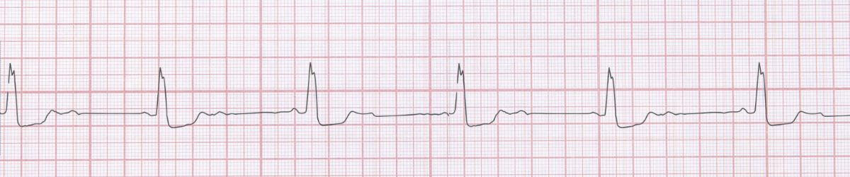
Figure 1: Second Degree AV Block Mobitz Type I - Characteristic ECG Pattern
Second Degree AV Block Mobitz Type I ECG Example
Figure 1: Second Degree AV Block Mobitz Type I - Characteristic ECG Pattern
🔑 Key Points at a Glance
- Heart Rate: Variable (60-100 bpm)
- Primary Significance: AV nodal level block, better prognosis than Mobitz II, often reversible
- Key Management: Observation if asymptomatic, rarely requires pacemaker
- Clinical Category: Clinical
Overview and Clinical Significance
Second Degree AV Block Mobitz Type I represents an important cardiac rhythm pattern that clinicians must accurately identify. AV nodal level block, better prognosis than Mobitz II, often reversible
Understanding this rhythm is essential for emergency physicians, cardiologists, intensivists, and all healthcare providers involved in acute cardiac care. Early recognition and appropriate management can significantly impact patient outcomes.
ECG Characteristics and Recognition
📊 Diagnostic ECG Criteria
- Progressive PR prolongation
- Dropped QRS after longest PR
- Wenckebach phenomenon
- Narrow QRS usually
Systematic ECG Analysis Approach
When analyzing any ECG, including suspected Second Degree AV Block Mobitz Type I, follow this systematic approach:
- Rate: Calculate the ventricular rate using the 300-150-100-75-60-50 rule or count complexes in 6 seconds × 10
- Rhythm: Assess regularity by measuring R-R intervals across the strip
- P Waves: Identify presence, morphology, and relationship to QRS complexes
- PR Interval: Measure from start of P wave to start of QRS (normal: 0.12-0.20 seconds)
- QRS Complex: Assess duration (normal: 1mm is significant)
- T Waves: Check morphology, direction, and concordance with QRS
- QT Interval: Measure and correct for heart rate (QTc normal: Correlate ECG findings with clinical presentation - the patient, not the monitor, determines management urgency
Evidence-Based Management
Acute Management Strategy
Primary Treatment Approach: Observation if asymptomatic, rarely requires pacemaker
Pharmacologic Interventions
Most cases require no pharmacologic intervention - focus on treating underlying causes and monitoring for progression.
Procedural Considerations
Procedural intervention is rarely required - conservative management is typically appropriate.
Differential Diagnosis
🔍 Consider These Mimics
- Mobitz II - no progressive PR prolongation
- Blocked PACs - look for premature P waves
- 2:1 AV block - cannot distinguish type without longer strip
Complications and Risk Stratification
Potential complications associated with Second Degree AV Block Mobitz Type I include:
- Progression to more severe conduction abnormalities
- Symptoms interfering with quality of life
Long-Term Management and Follow-Up
Regular outpatient follow-up with cardiology or electrophysiology is recommended to monitor for progression and optimize therapy.
📅 Follow-Up Recommendations
- Cardiology follow-up within 2-4 weeks
- Consider Holter monitor or event recorder for recurrent symptoms
- Lifestyle modifications: exercise, stress reduction, avoid triggers
Common Pitfalls and How to Avoid Them
⚠️ Common Mistakes to Avoid
- Failing to correlate ECG with clinical presentation
- Missing underlying reversible causes
- Not consulting cardiology when uncertain
Patient Education and Counseling
When counseling patients diagnosed with Second Degree AV Block Mobitz Type I, address the following key points:
- Nature of the condition: Explain the rhythm abnormality in simple terms, avoiding medical jargon
- Prognosis: Provide realistic expectations about symptom control and quality of life
- Warning signs: Educate about symptoms requiring immediate medical attention (chest pain, syncope, severe dyspnea)
- Medication compliance: Importance of taking prescribed medications as directed
- Lifestyle modifications: Limit caffeine and alcohol, maintain healthy weight, exercise regularly (as tolerated), stress reduction
- Activity restrictions: Generally no restrictions once symptoms controlled
Evidence-Based Guidelines and References
Current management of Second Degree AV Block Mobitz Type I is based on evidence from major clinical trials and consensus guidelines from professional societies including:
- American Heart Association (AHA) / American College of Cardiology (ACC) Guidelines
- European Society of Cardiology (ESC) Guidelines
- Advanced Cardiac Life Support (ACLS) Protocols
- Heart Rhythm Society (HRS) Expert Consensus Statements
📚 Level of Evidence
Most recommendations for acute management of Second Degree AV Block Mobitz Type I are supported by Level B (limited randomized trials or observational studies) evidence.
Summary and Clinical Bottom Line
📋 Clinical Bottom Line
Second Degree AV Block Mobitz Type I is characterized by progressive pr prolongation and dropped qrs after longest pr. AV nodal level block, better prognosis than Mobitz II, often reversible Management priority: Observation if asymptomatic, rarely requires pacemaker Key takeaway: Prompt diagnosis and appropriate therapy optimize outcomes
About the Author
Dr. Raj K
Emergency Medicine Physician Dr. Raj K is a board-certified Emergency Medicine physician with extensive experience in acute cardiac care and ECG interpretation. He is passionate about medical education and bringing evidence-based emergency medicine knowledge to healthcare providers worldwide through E-PulsePoints.