 Ventricular Tachycardia ECG Example
Figure 1: Ventricular Tachycardia - Characteristic ECG Pattern
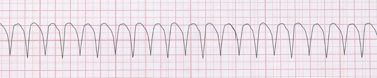
Ventricular Tachycardia ECG Example
Figure 1: Ventricular Tachycardia - Characteristic ECG Pattern
🔑 Key Points at a Glance
- Heart Rate: >100 bpm (typically 140-200)
- Primary Significance: Life-threatening arrhythmia, can degenerate to VFib, may indicate structural heart disease
- Key Management: If stable: amiodarone or procainamide. If unstable: synchronized cardioversion. Pulseless: defibrillation
- Clinical Category: Clinical
Overview and Clinical Significance
Ventricular Tachycardia represents a critical cardiac rhythm pattern that requires immediate recognition and intervention. Life-threatening arrhythmia, can degenerate to VFib, may indicate structural heart disease
Understanding this rhythm is essential for emergency physicians, cardiologists, intensivists, and all healthcare providers involved in acute cardiac care. Early recognition and appropriate management can significantly impact patient outcomes.
ECG Characteristics and Recognition
📊 Diagnostic ECG Criteria
- Wide QRS complexes >0.12s
- Regular or slightly irregular
- AV dissociation
- Capture or fusion beats
Systematic ECG Analysis Approach
When analyzing any ECG, including suspected Ventricular Tachycardia, follow this systematic approach:
- Rate: Calculate the ventricular rate using the 300-150-100-75-60-50 rule or count complexes in 6 seconds × 10
- Rhythm: Assess regularity by measuring R-R intervals across the strip
- P Waves: Identify presence, morphology, and relationship to QRS complexes
- PR Interval: Measure from start of P wave to start of QRS (normal: 0.12-0.20 seconds)
- QRS Complex: Assess duration (normal: 1mm is significant)
- T Waves: Check morphology, direction, and concordance with QRS
- QT Interval: Measure and correct for heart rate (QTc normal: Always assess hemodynamic stability before initiating treatment - unstable patients require immediate intervention regardless of the specific arrhythmia
Evidence-Based Management
Acute Management Strategy
Primary Treatment Approach: If stable: amiodarone or procainamide. If unstable: synchronized cardioversion. Pulseless: defibrillation
🚨 Emergency Protocol
- Assess ABC (Airway, Breathing, Circulation) immediately
- Attach cardiac monitor and obtain 12-lead ECG
- Establish IV access and administer oxygen if SpO₂ Most recommendations for acute management of Ventricular Tachycardia are supported by Level A (multiple randomized trials) evidence.
Summary and Clinical Bottom Line
📋 Clinical Bottom Line
Ventricular Tachycardia is characterized by wide qrs complexes >0.12s and regular or slightly irregular. Life-threatening arrhythmia, can degenerate to VFib, may indicate structural heart disease Management priority: If stable: amiodarone or procainamide. If unstable: synchronized cardioversion. Pulseless: defibrillation Key takeaway: Immediate recognition and treatment are critical for patient survival
About the Author
Dr. Raj K
Emergency Medicine Physician Dr. Raj K is a board-certified Emergency Medicine physician with extensive experience in acute cardiac care and ECG interpretation. He is passionate about medical education and bringing evidence-based emergency medicine knowledge to healthcare providers worldwide through E-PulsePoints.